
[Posselt clinical cases]
Restoring right back teeth with implants and sinus grafting
Before and after, side by side.
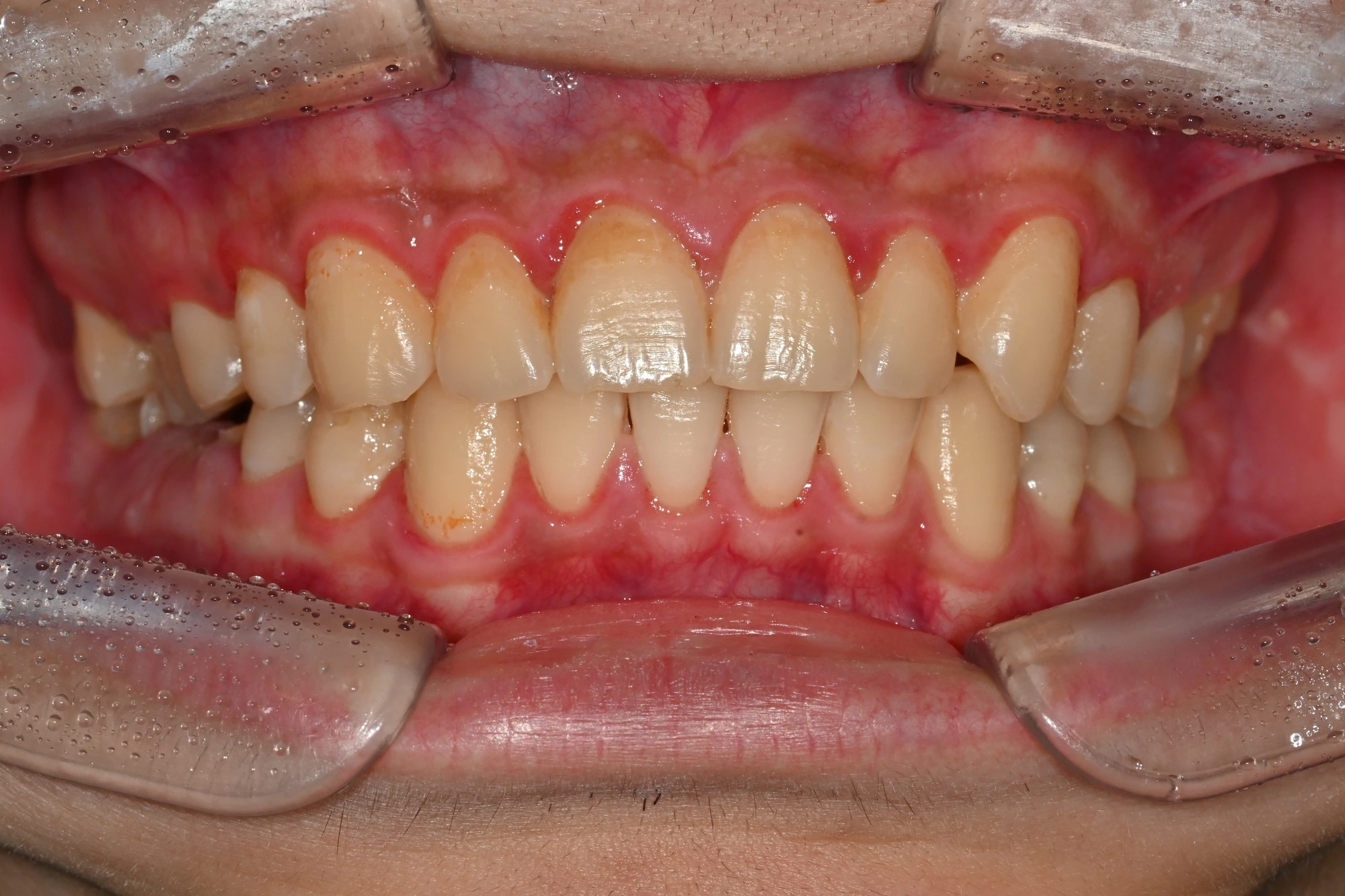
Panoramic radiographs: before and after
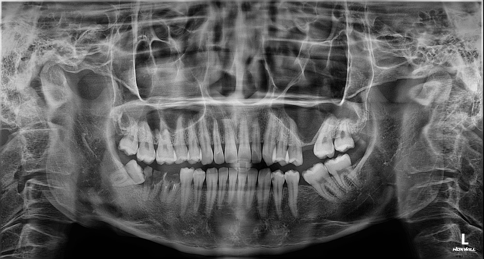
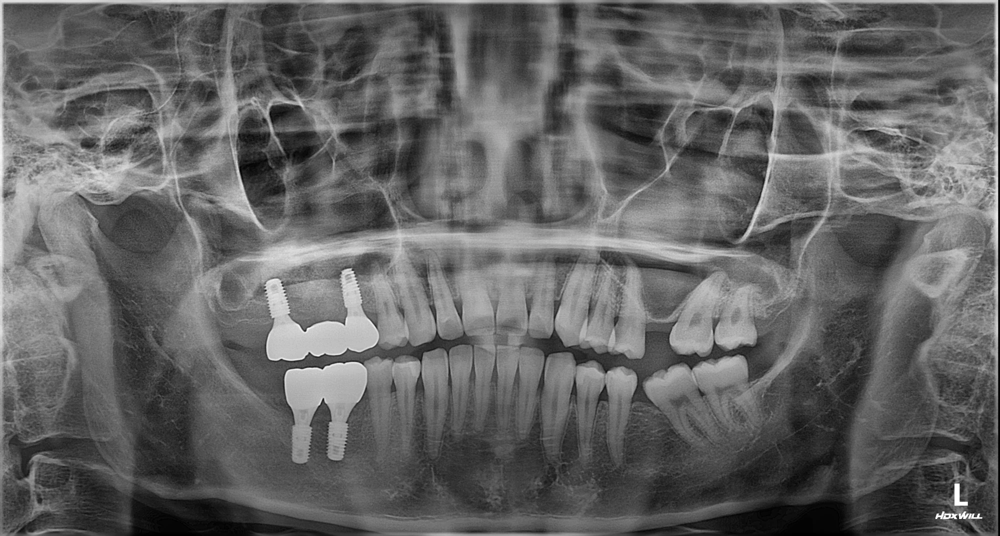
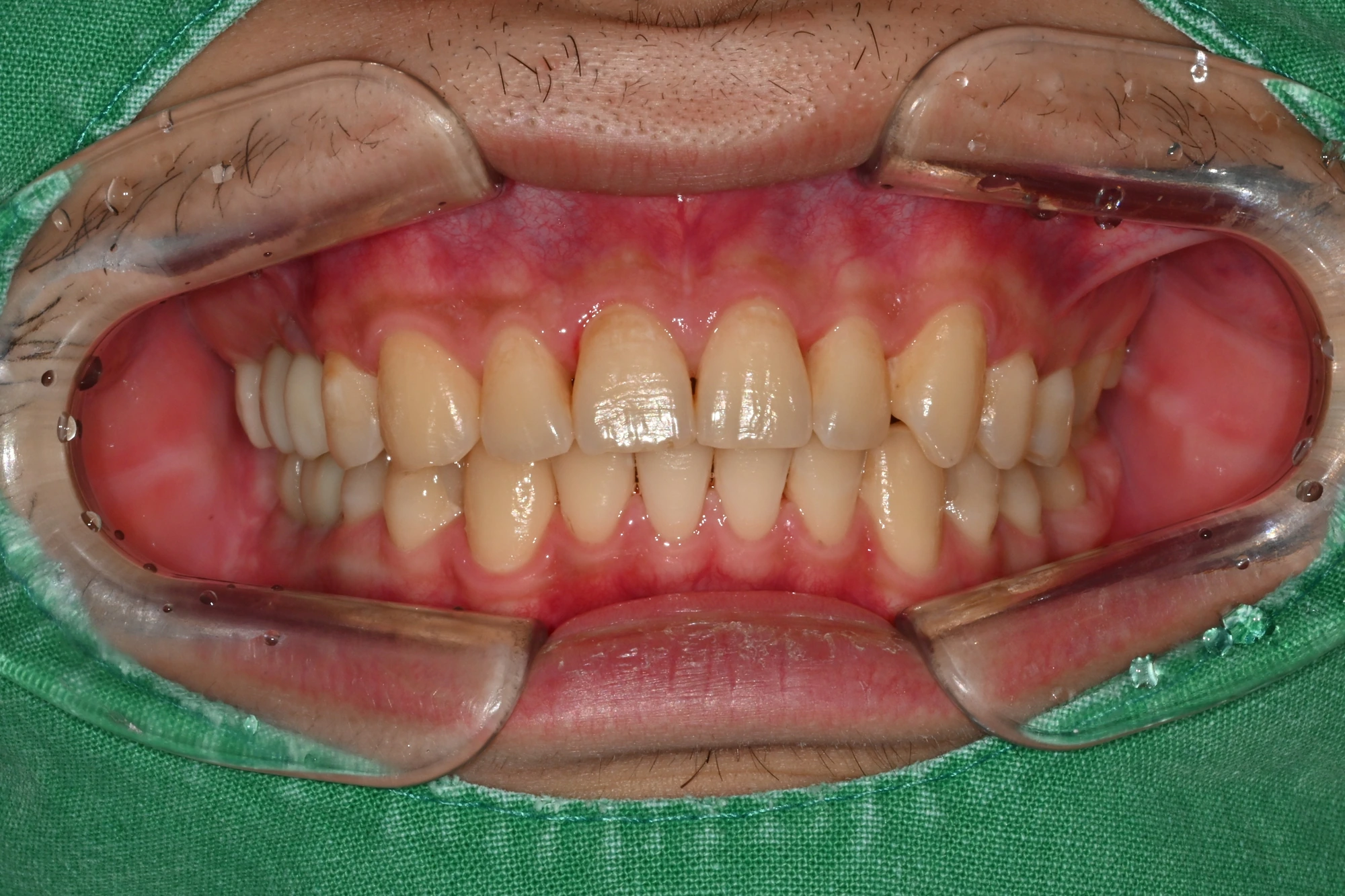
Actual records of the same patient. Capture dates, angles and lighting differ.
About this treatment
After tooth loss, opposing teeth had overerupted and neighbouring teeth had tilted mesially, altering the bite. Treatment included extraction, upper-right bone grafting through a lateral approach, and staged implant-supported restoration of the upper and lower right posterior teeth.
Treatment journey
Photographs are arranged chronologically. Intervals refer to the photographic record, not a confirmed treatment duration or a guaranteed outcome.
Before treatment
The pretreatment photographs show missing teeth and the condition of the remaining teeth.
Before treatment · Frontal view
View full-size photo ↗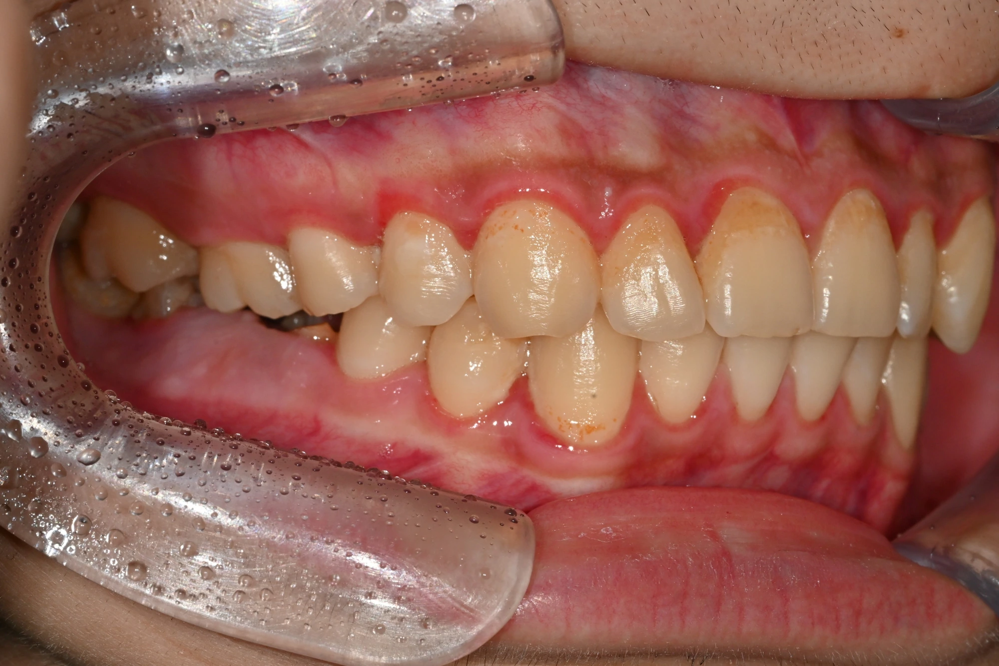
Before treatment · Right buccal view
- Right posterior gap and bite
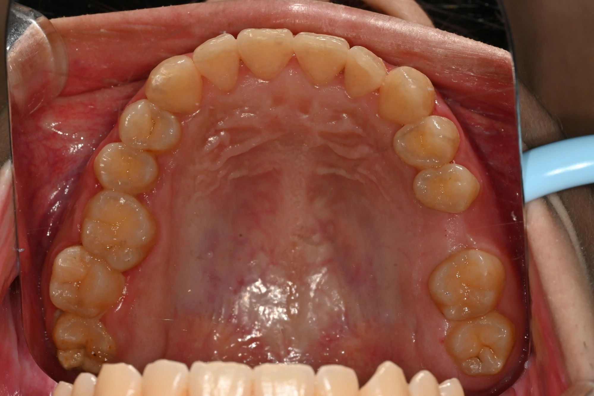
Before treatment · Upper occlusal view
View full-size photo ↗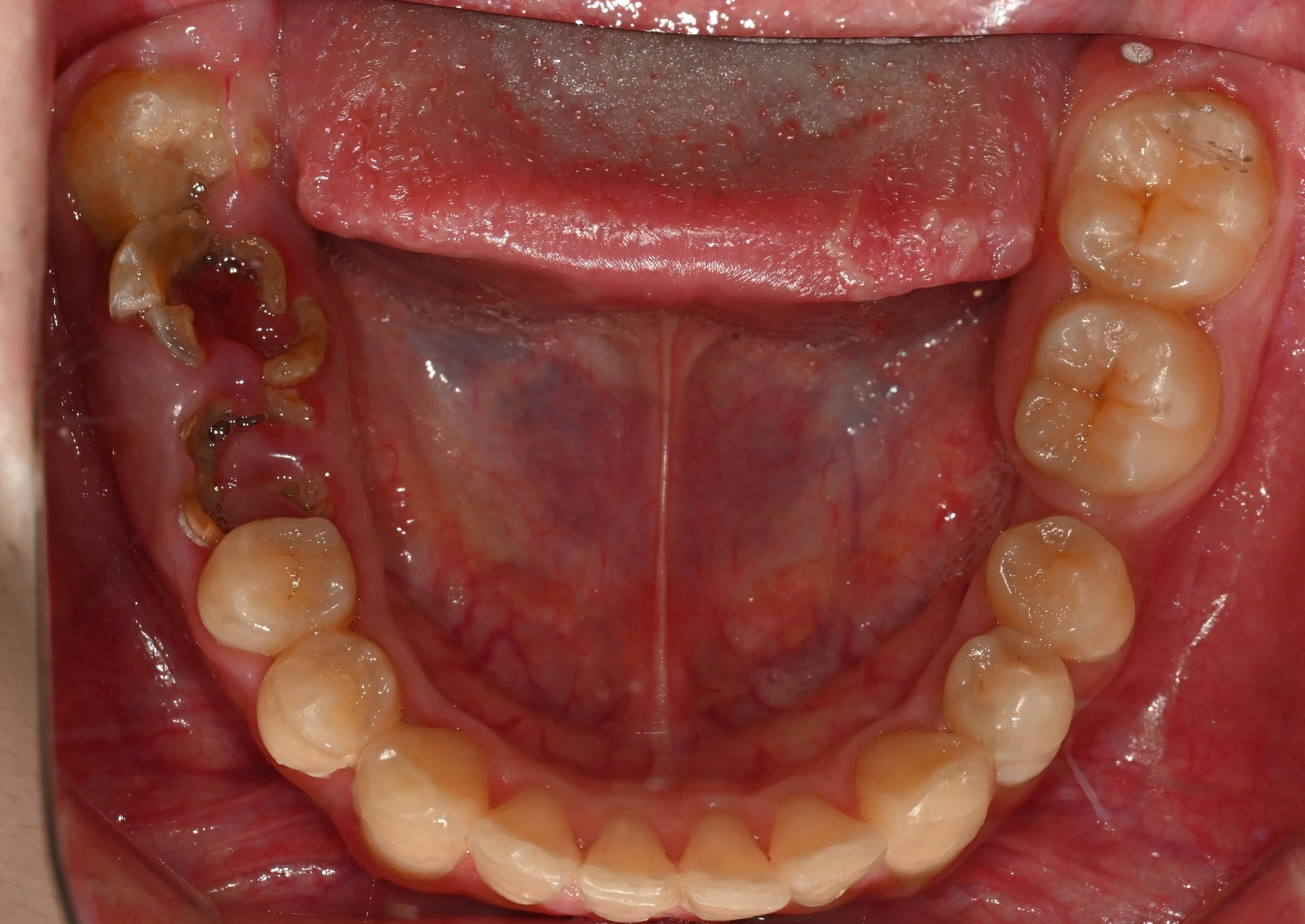
Before treatment · Lower occlusal view
View full-size photo ↗Before treatment · Panoramic radiograph
- Upper-right posterior position
- Missing lower-right posterior teeth
Extraction, grafting and healing
These records show the panoramic view after lower-right treatment, the sutured upper-right graft site, and follow-up radiographs.
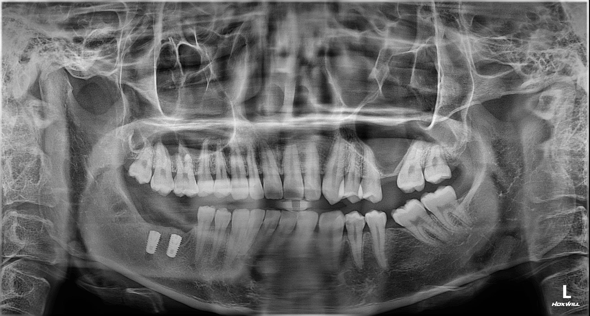
After treatment in the lower right · Panoramic radiograph
View full-size photo ↗Show postoperative suture photograph
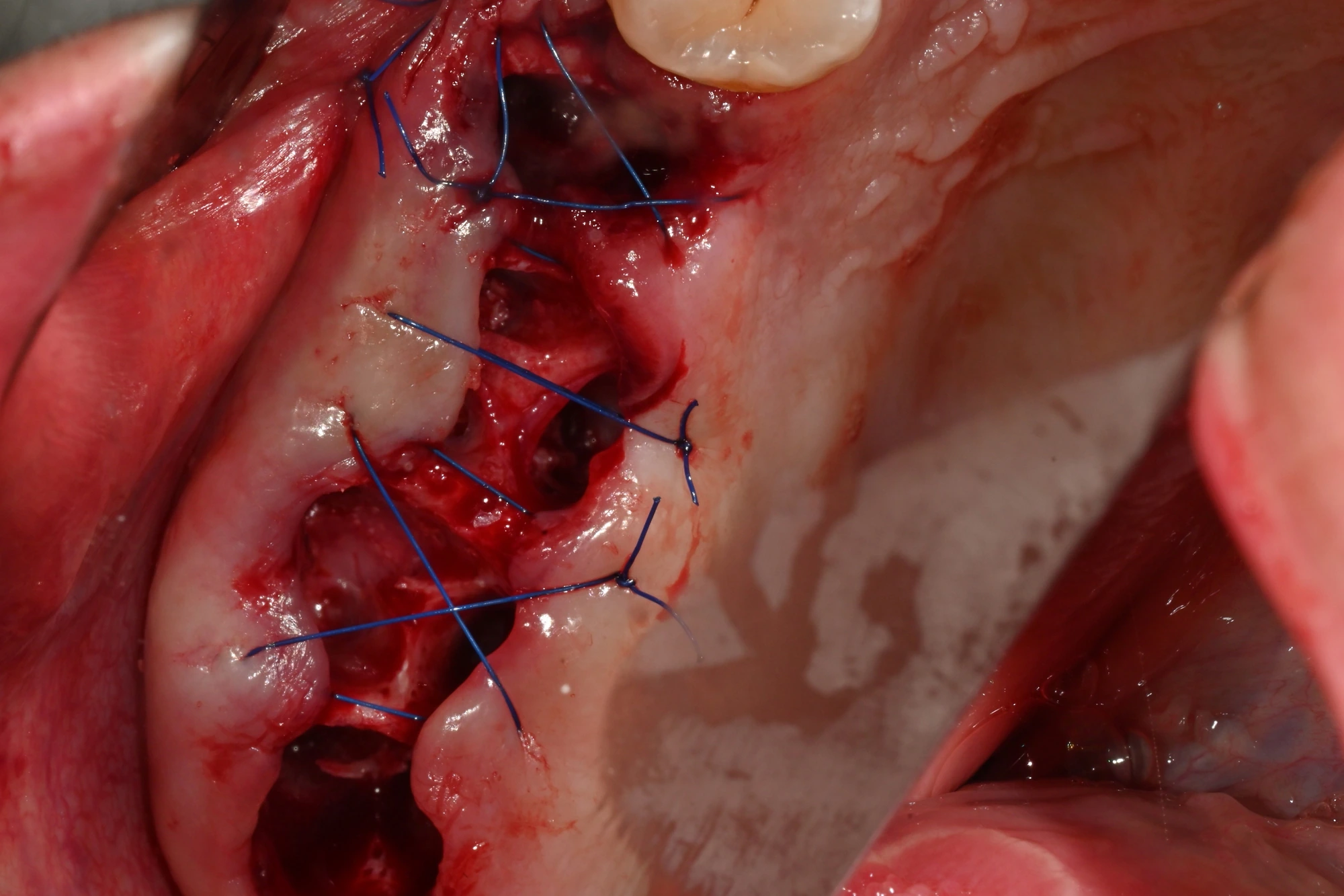
About 3 weeks later · After upper-right bone grafting · Sutured area
View full-size photo ↗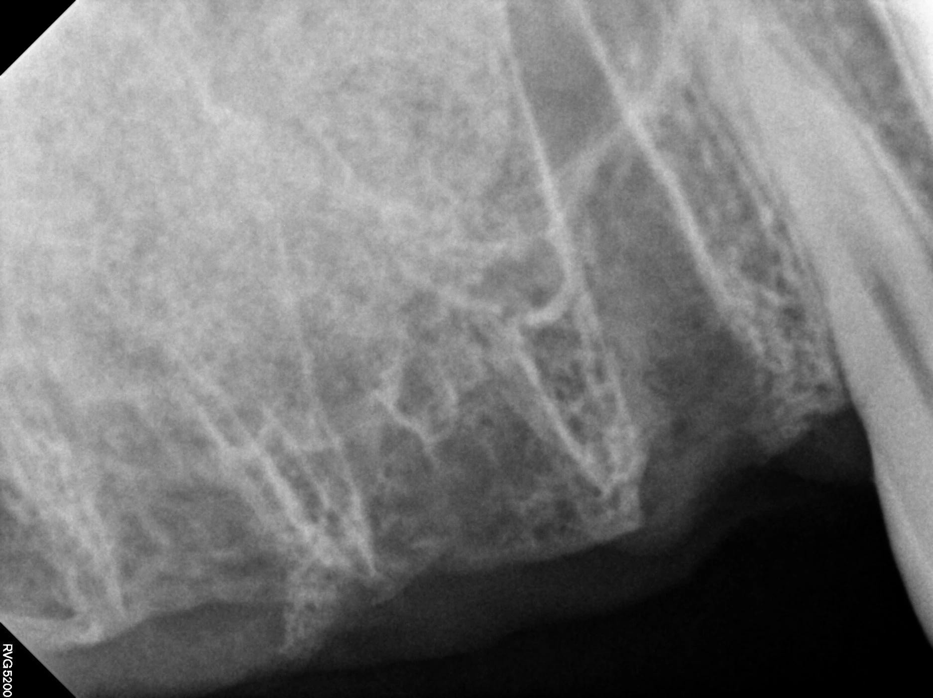
About 3 weeks later · Follow-up after bone grafting · Periapical radiograph
View full-size photo ↗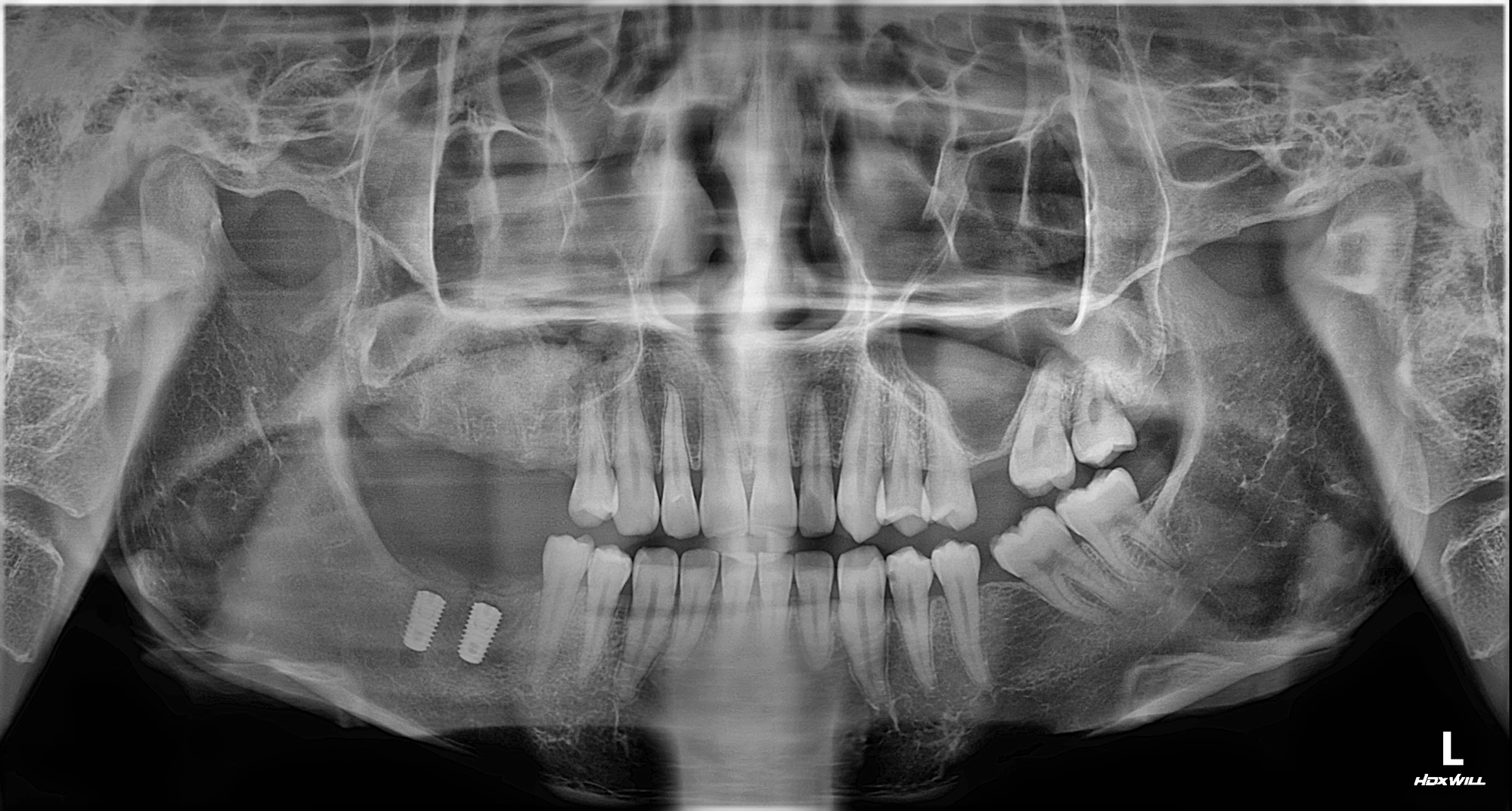
About 1 month later · Follow-up after bone grafting · Panoramic radiograph
View full-size photo ↗Upper implants and restorative preparation
Panoramic radiographs before and after upper-right implant placement are followed by the healing-abutment stage. Healing abutments are used during recovery before the final teeth are fitted.
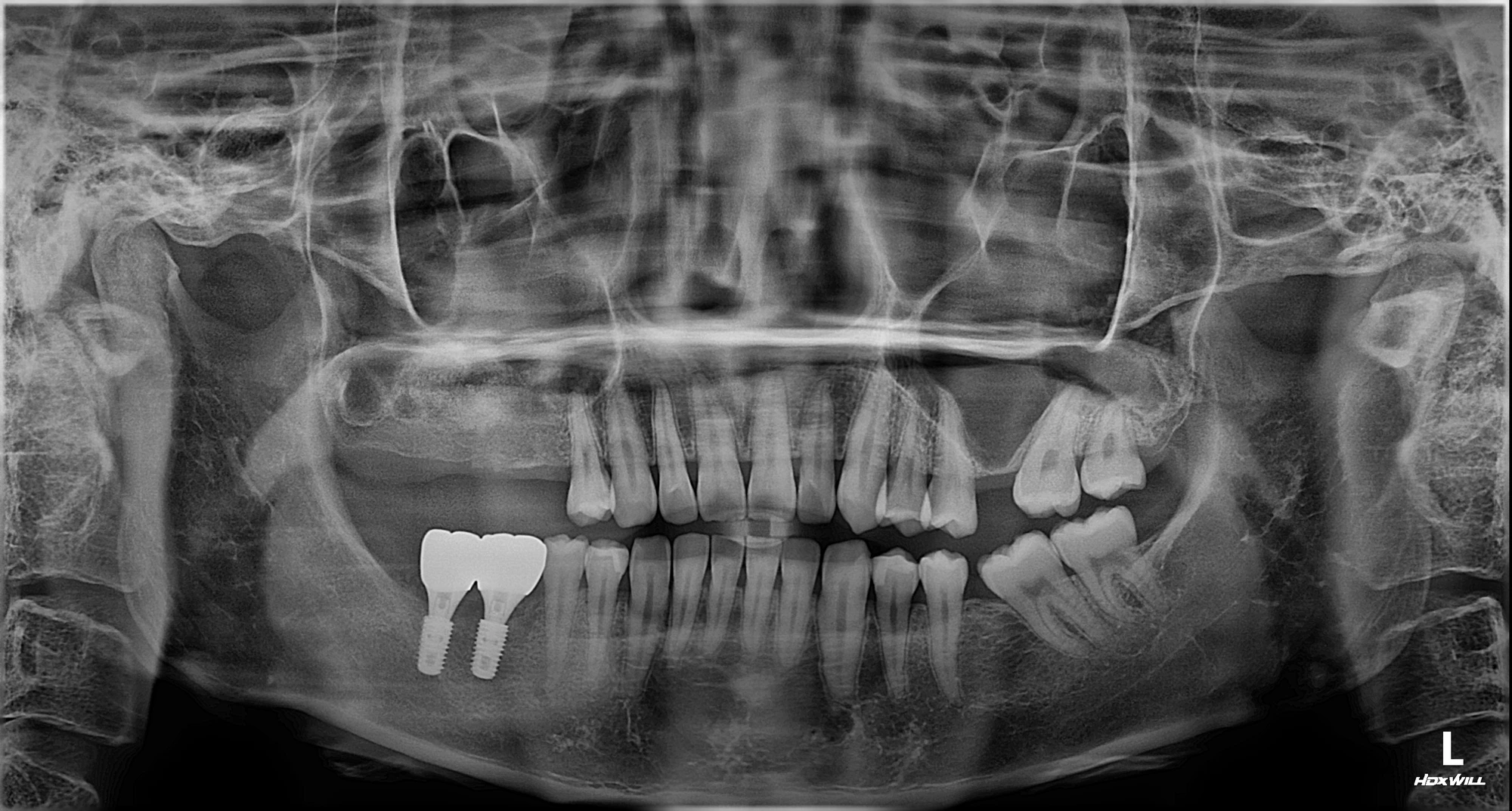
About 6 months later · Before upper-jaw implant placement · Panoramic radiograph
- Upper-right graft follow-up area
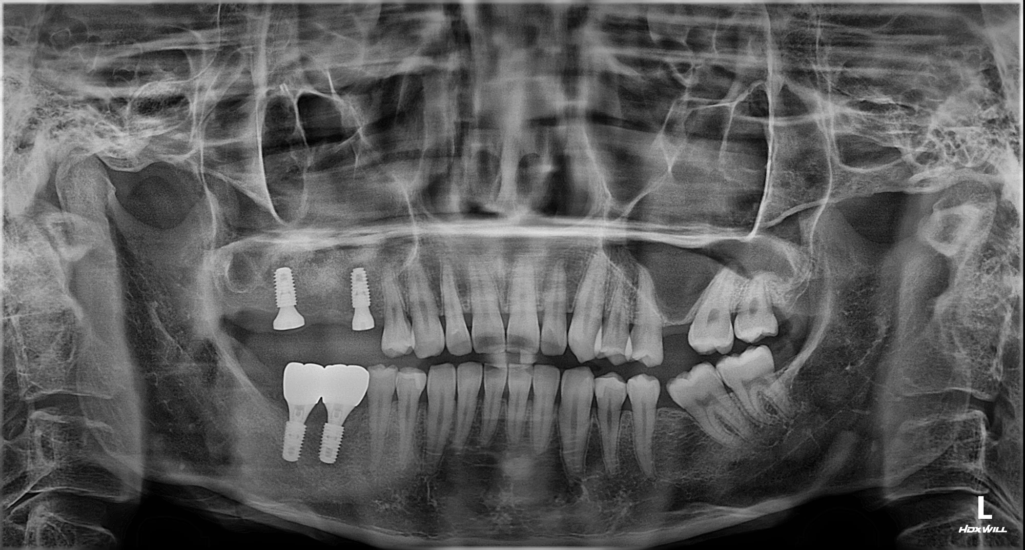
About 6 months later · After upper-right implant placement · Panoramic radiograph
View full-size photo ↗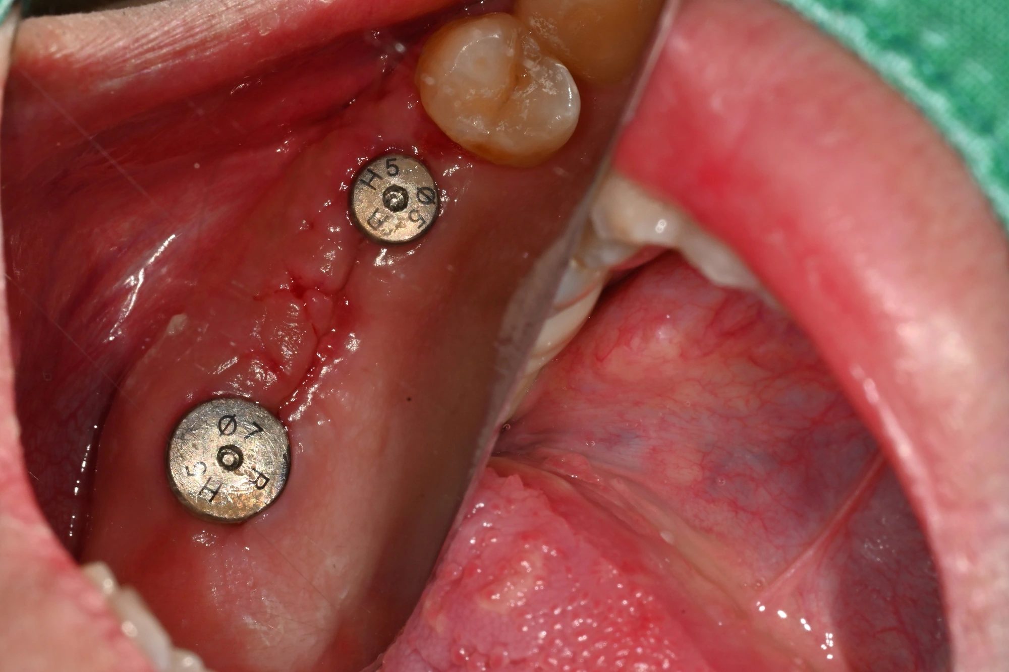
About 6 months later · Upper right · Healing-abutment stage
View full-size photo ↗Restoration and later follow-up
The radiograph with restorations is followed by later front, side and occlusal photographs. Later follow-up intervals are measured from that panoramic record.
After restoration placement · Panoramic radiograph
View full-size photo ↗About 7 months later · Follow-up after restoration placement · Frontal view
View full-size photo ↗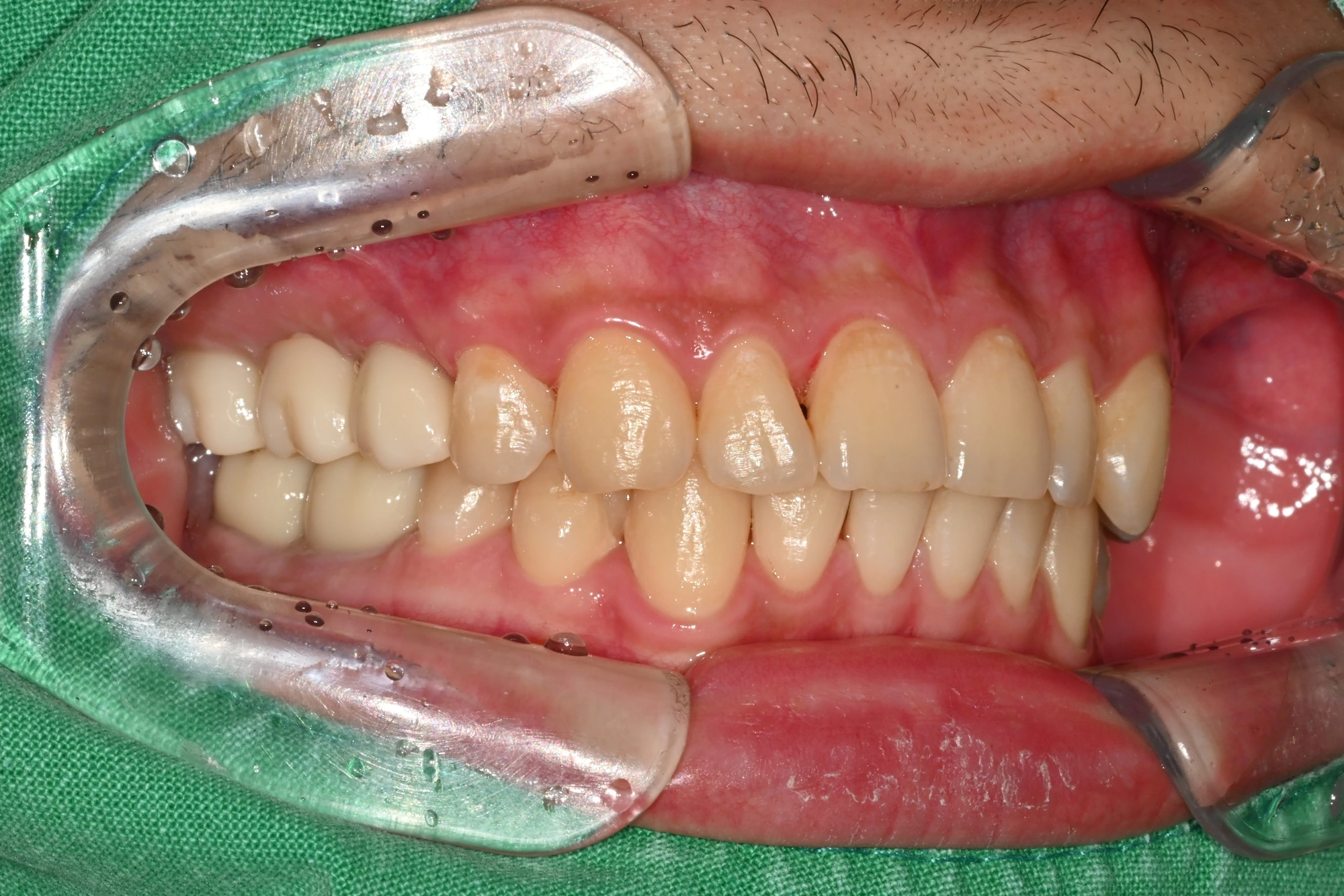
About 7 months later · Follow-up after restoration placement · Right buccal view
View full-size photo ↗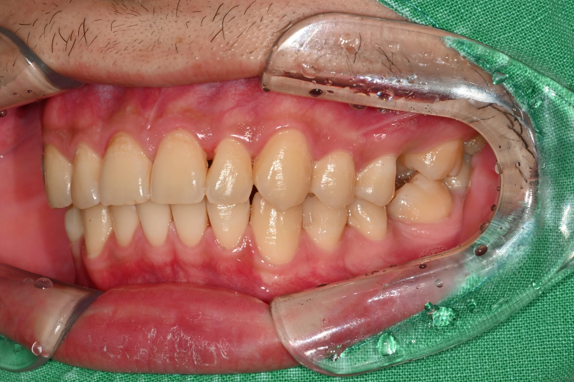
About 7 months later · Follow-up after restoration placement · Left buccal view
View full-size photo ↗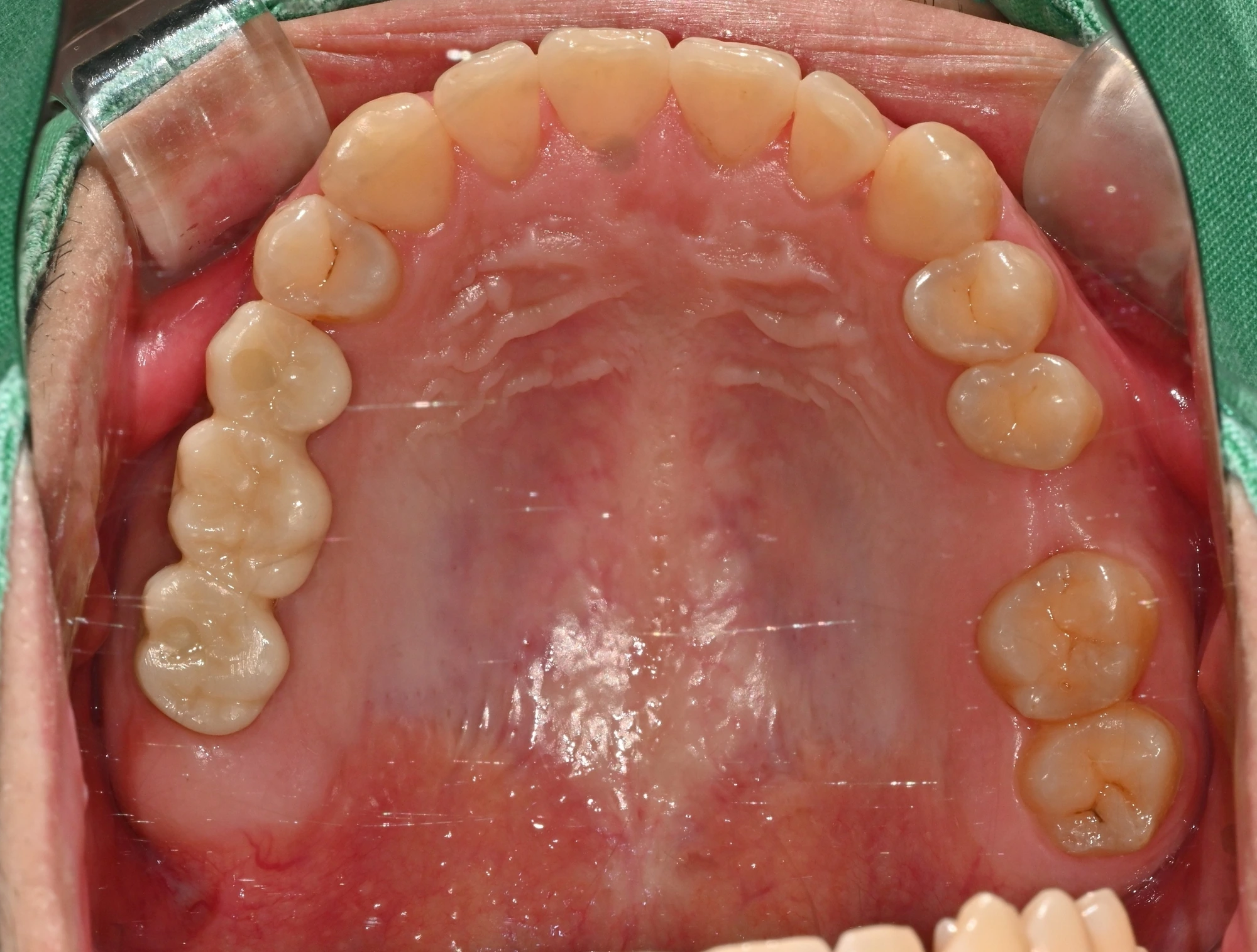
About 7 months later · Follow-up after restoration placement · Upper occlusal view
- Upper-right restoration follow-up
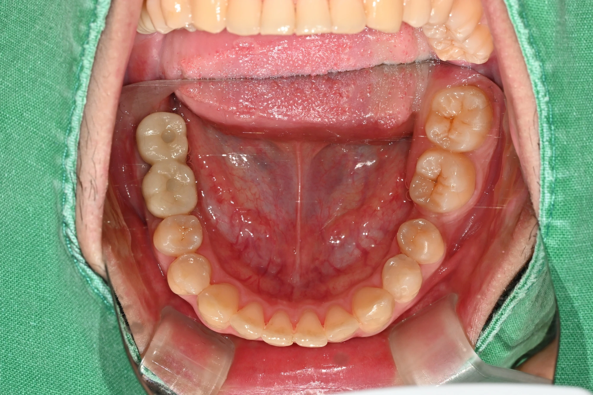
About 7 months later · Follow-up after restoration placement · Lower occlusal view
- Lower-right restoration follow-up
Implant treatment carries risks, including infection, peri-implant inflammation and damage to the restoration. Treatment, healing time and outcomes vary from person to person.
Ask about posterior teeth and bone grafting
